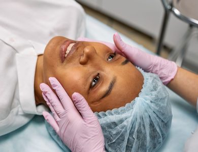
Why “Before and After” Means More Than Just Swollen Cheeks
Most sinus lift galleries show two images: a pre-op CT scan with barely any bone, and a pristine post-op x-ray months later. What’s missing is everything in between—the bruising, the uneven smile, the week where you wonder if your face will ever look normal again. That gap fuels the anxiety that keeps people second-guessing a medically necessary procedure.
A true “before and after” unfolds across three distinct layers.
The radiographic layer is what your surgeon tracks. On a CBCT scan, a healthy graft shows a dense, well-contained bone mass increasing vertical height by roughly 8–12 mm. The facial aesthetic layer is what you’ll track daily: swelling that peaks around day 3, faint bruising that can drift below the eye, and a smile that might feel stiff or asymmetrical for two to three weeks. None of this means the graft is failing. It means your body is doing exactly what the Mayo Clinic describes as a normal inflammatory healing response—forming a blood clot scaffold that precursor cells slowly remodel into living bone over four to nine months.
The functional layer catches people off guard. Expect temporary sinus pressure, a sensation of fullness, and a nasal tone to your voice for the first few weeks. What you shouldn’t expect is sharp pain, fever, or loose graft particles migrating into your nose—those are red flags that warrant an immediate call to your surgeon.
A sinus lift is never the final “after.” It’s a foundation surgery. The real endpoint isn’t a healed graft on a scan—it’s the moment an implant crown is seated in that new bone. The graft is the basement. The smile is the finish line.
Reading the Scans: Bone Height Before Grafting vs. After Maturation
If you felt a flash of dread when your dentist pulled up your CT scan and pointed to a ghostly gray void above your back teeth, you’re not alone. That transparent, air-filled space is your maxillary sinus, and in a healthy jaw, a thick floor of bone should separate it from your tooth roots. When that bone measures less than 4–6 mm in height—roughly the thickness of two stacked dimes—a dental implant has no structural anchorage. The sinus membrane sags low into the space where bone should be, making it impossible to place an implant without puncturing it during drilling. This is the “before” image that triggered your surgical referral.
The immediate post-operative scan tells a different story. Once the sinus membrane has been gently lifted and bone graft material is packed into the newly created space, the imaging shifts from hollow to hyper-dense. Radiopaque graft particles appear as a bright, cloud-like mass forming a distinct, dome-shaped floor where the sinus once dipped low. This “lifted” architecture isn’t load-bearing yet; it’s a biological scaffold. According to the American Academy of Periodontology, the graft serves as a framework for your own bone cells to migrate into, a process that takes months, not weeks.
By the 6- to 9-month mark, a successful maturation scan reveals the payoff. That bright, particulate cloud resolves into uniform, cortical bone density with no visible gaps or granular separation. The new sinus floor looks like a smooth, continuous shelf of solid bone—typically 10–15 mm in vertical height. For your surgeon, this is the green light for implant placement: a stable, vascularized platform that can withstand the torque of a titanium post. If the density looks patchy or voids persist, the implant timeline gets pushed back, not cancelled—but seeing that uniform white band on the screen is the moment the waiting finally translates into a viable foundation.
The First 72 Hours: Normal Facial Swelling vs. Warning Signs
The face looking back at you in the mirror 48 hours after surgery is not the final result—it’s a temporary, fluid-filled version of you that can look genuinely alarming if you weren’t expecting it. Swelling follows a predictable arc: mild puffiness on day one, a dramatic peak around 36 to 48 hours, then a slow, steady deflation through day three. Because the sinus membrane sits just below the eye, fluid often tracks downward with gravity, creating a crescent-shaped bruise that can spread from the upper cheek to the lower eyelid. That yellowish-green fade that appears by day four or five is a sign of normal hemoglobin breakdown, not infection.
One of the most common panic triggers is asymmetry. It is completely normal for the side where the surgeon accessed the sinus to swell significantly more. If your procedure was on the right side, your right cheek and upper lip may look inflated while the left side remains relatively unchanged. This is not a hematoma or a failed graft—it’s simply the path of least resistance for post-surgical fluid in the tissue planes.
What you cannot ignore are signs that the inflammatory response has tipped into something pathological. According to the American Association of Oral and Maxillofacial Surgeons, a rapidly expanding swelling that feels shiny, taut, and hot to the touch—especially when accompanied by fever—warrants an immediate call. Inside your mouth, normal bruising on the cheek side of the gums is expected, but deep purple or black discoloration spreading across the palate is not; that pattern can signal a palatal hematoma or compromised blood flow to the graft site. Any persistent foul odor, salty taste, or visible pus seeping from the incision line are classic indicators that bacteria have colonized the surgical site, and oral antibiotics may need to start within hours to salvage the graft.
How Your Smile and Lip Position Change During Healing
One of the quiet fears nobody voices in the consult room is whether you’ll look in the mirror during the months between surgery and implants and see a stranger. Here’s what actually happens to your smile and lip position—and why the changes are almost always temporary.
The initial fullness you see in the first 10–14 days is surgical edema, and it will exaggerate the upper lip and cheek on the grafted side. You might notice the nasolabial fold—the line from your nose to the corner of your mouth—looks softer or slightly “filled.” A Journal of Oral and Maxillofacial Surgery review notes that transient midface swelling after sinus augmentation peaks around day 3 and resolves enough by day 14 that patients can reliably see their underlying facial contours again.
Once the fluid recedes, the graft material itself can leave a subtle, firm contour along the cheek-side bone. For grafts placed in the posterior maxilla (molars only), this is far behind the smile curtain. Your lip position, tooth display, and smile line won’t change in any way you or anyone else would notice. The smile zone is an anterior structure, and a molar sinus lift simply doesn’t reach it.
The nuance comes with premolar or canine-area grafts. Here, a buccal graft can provide mild structural support to the upper lip during the edentulous phase, especially if you’ve had significant bone loss. Some patients describe it as looking like they have a thin, invisible retainer behind the lip—slightly more “lifted” than the opposite side. This is a contour change, not a functional impairment, and it typically softens as the graft remodels over 4–6 months. By the time your implant crown is seated, the prosthetic itself restores natural lip support, and any residual graft volume simply reads as healthy facial structure.
The 2-Week to 3-Month Window: When the Face Settles but the Graft Is Fragile
Here’s the cruel trick of the sinus lift recovery timeline: The moment you finally look like yourself again is the moment your graft is at its most vulnerable. By the end of week two, the purple and yellow bruising under your eye has usually faded to a faint shadow, and your cheek contour has softened back to its normal shape. You’ll catch your reflection and think, “Okay, I’m through it.” That external healing masks a critical internal reality. The bone graft particles are still loose, unintegrated granules at this stage—think wet sand packed into a cavity, not solid structure.
The Confidence Gap That Causes Graft Failure
This disconnect between how you feel and what’s actually happening inside your sinus is what surgeons call the “confidence gap,” and it’s responsible for a significant share of early graft failures. You feel fine, so you bend down to pick up a heavy grocery bag, or you instinctively blow your nose with force during allergy season. That sudden pressure change can tear the membrane or physically displace the graft particles. Strict activity restrictions for at least 3–4 weeks are non-negotiable, but many patients abandon them the moment external swelling drops. Heavy lifting, resistance training, playing a wind instrument, or even stifling a violent sneeze by pinching your nose closed can generate enough intra-sinus pressure to compromise the entire site.
Red Flags: What a Failing Graft Feels Like in This Window
A healthy graft site at this stage often feels firm or slightly “woody” under the cheek, not soft. That’s normal—woven bone hasn’t matured yet. What isn’t normal is a sudden deflation of the cheek contour, or the sensation of gritty granules migrating down into your mouth or out of your nostril. If you notice a persistent salty or metallic taste, or if your sinus congestion suddenly includes small, sand-like particles when you gently rinse, contact your surgeon immediately. These are signs the graft is being expelled. A minor sinus infection might respond to antibiotics, but particulate matter in your mucus almost always signals mechanical displacement. Catching it within the first 48 hours can mean the difference between a quick patch procedure and starting the entire graft from scratch after months of wasted healing time.
Functional Changes: Breathing, Sinus Pressure, and Nasal Tone
During the first week, the “heavy sinus” sensation is real, but it’s driven by temporary post-surgical edema and the physical presence of graft material pushing gently against the sinus membrane. Your sinus isn’t blocked with cement; it’s swollen tissue reacting to a new neighbor. Congestion, a feeling of fullness, and even some blood-tinged nasal discharge are expected parts of the inflammatory phase, not signs that the graft failed.
That early pressure fades as the swelling resolves, usually between days 5 and 10. Long-term, the sinus cavity does end up slightly smaller—a few millimeters of vertical bone gain directly reduces the air space above it. But here’s what matters functionally: the cilia lining your sinuses don’t quit. According to the American Rhinologic Society, healthy ciliary function and natural drainage pathways are remarkably adaptive. Once the membrane heals and any minor inflammation settles, mucus clearance typically normalizes. Most patients report zero chronic change in how they breathe through their nose, and there’s no evidence that a standard sinus lift triggers new allergy problems or chronic sinusitis in otherwise healthy cavities.
The voice change fear is largely overblown. Your sinuses do contribute to vocal resonance, but a unilateral graft or a modest bilateral lift changes the volume of the space so subtly that it’s acoustically imperceptible to listeners. Minor resonance shifts can occur with very large bilateral grafts that significantly reduce both sinus cavities, but even then, the difference is usually noticeable only to the patient—like the slight vocal fatigue you might feel with a mild cold. It won’t alter your speaking voice enough for anyone else to ask if you’re sick.
The Final Picture: Implant Crowns Over Grafted Bone
When you finally see the crown emerge from the gum tissue, the entire surgical timeline suddenly makes sense. The “before” image that likely lives in your chart—a collapsed posterior bite with a sunken upper lip and a black triangle where a molar used to be—looks like a different person. The “after” isn’t a tooth; it’s the restoration of vertical dimension, the invisible height that supports your lips and cheeks. Without the sinus lift creating a solid bony floor, that crown would have nowhere to anchor except thin air.
Adequate bone volume does something most patients don’t anticipate: it lets the dental lab design a proper emergence profile. When bone is deficient, technicians are forced to compensate with a longer clinical crown or a strip of pink porcelain meant to mimic gum tissue, which the American Academy of Periodontology notes can compromise long-term peri-implant health by creating non-cleansable contours. With 8–10 mm of regenerated bone height, the crown emerges from the tissue at the same angle and proportion as a natural tooth. No pink ceramic camouflage. No “horse tooth” elongation.
The soft tissue tells the real success story. A flat, knife-edged ridge with intact interdental papillae—those tiny triangles of gum filling the spaces between teeth—is the visual hallmark of graft integration. If the papillae are present and the ridge doesn’t bulge, you’re looking at proof that the bone maintained its volume during healing. That architecture also acts as a biological seal, keeping bacteria out and preserving the aesthetic result for years rather than months.
How to Verify Your Surgeon’s Graft Success Rate Before You Commit
Most surgeons will happily show you a binder of perfect implant smiles, but that tells you almost nothing about how they handle the cases that actually resemble yours. You need to see the data that lives one layer deeper—the bone graft integration, not the crown on top.
The CBCT Audit: Ask for Their Scans, Not a Stock Photo
A reputable surgeon can pull up before-and-after cone-beam computed tomography (CBCT) scans from their own archive that match your specific bone deficiency—typically a subantral height of less than 4–6 mm. You’re looking for the actual gray-and-black cross-sections showing the sinus floor lifted and the graft material consolidated into dense, vascularized bone after 6–9 months of healing. If they reach for a dental implant manufacturer’s marketing brochure instead, you’re looking at curated aspirations, not surgical outcomes.
The Perforation Rate Question That Separates Candor from Salesmanship
Schneiderian membrane perforation is the most common intraoperative complication, and every surgeon tears one eventually. The difference is whether they’ll tell you their personal rate. Ask directly: “What percentage of your sinus lifts involve a membrane tear, and how do you manage it when it happens?” An honest answer typically falls in the 10–30% range, depending on case complexity, followed by a clear protocol—usually a resorbable collagen membrane patch, possibly a brief pause in the procedure, and rarely any impact on final implant success if repaired correctly. A surgeon who claims “it never happens” or dismisses the question is giving you a reason to keep interviewing.
Graft Material Logic: Why This Choice Isn’t Generic
The source of your graft material shapes both the healing timeline and the risk profile. Autogenous bone—harvested from your own mandible or iliac crest—offers the gold standard for osteogenic potential but requires a second surgical site. Allografts from accredited tissue banks eliminate donor-site morbidity and currently account for a substantial share of U.S. sinus augmentation procedures, while xenografts (typically bovine-derived) provide a slow-resorption scaffold that some surgeons prefer for maintaining volume in pneumatized sinuses. The key isn’t which material is “best” in the abstract—it’s whether your surgeon can explain why their choice fits your sinus anatomy, residual bone density, and implant timeline without defaulting to whatever happens to be on their shelf.
When to Seek Emergency Care: Signs Your Graft Is Compromised
Most post-operative discomfort follows a predictable, fading arc. But there’s a small cluster of symptoms that don’t belong on that curve—and waiting too long to act on them can turn a salvageable graft into a total loss.
The Salty Drip That Won’t Quit
A few drops of blood-tinged fluid from the nostril on the surgical side are normal in the first 48 hours. What’s not normal is a sudden, persistent salty discharge—often described as tasting like ocean water or broth—that continues past the 24-hour mark. This almost always signals a Schneiderian membrane perforation that has allowed graft particles to seep into the sinus cavity. When leakage goes unrecognized post-operatively, the infection risk climbs sharply. If you’re swallowing something salty every time you tilt your head forward, call your surgeon immediately.
One-Sided Pressure, Fever, and Foul Odor
Mild, diffuse congestion is expected. Severe, unilateral sinus pain—especially when paired with a fever above 100.4°F and a foul smell that you can’t rinse away—points toward acute graft infection or purulent sinusitis. This isn’t the same as the dull ache of healing bone; it’s a deep, throbbing pressure that often worsens when you bend over. Left untreated, an infected graft can necrose, requiring complete removal and delaying your implant timeline by 4–6 months.
Granules Where They Shouldn’t Be
If you notice gritty, sand-like particles visibly migrating through the gum incision or feel them exiting your nose, the graft site has lost containment. This is a mechanical failure that demands immediate surgical re-evaluation, not a wait-and-see approach. Document what you see, avoid blowing your nose entirely, and contact your surgical team within hours, not days.
The Emotional Timeline: When Patients Regret It and When They Don’t
Most patients hit a wall around Day 4. The swelling peaks, your upper lip feels stiff and foreign, and the mirror shows a face that doesn’t look like yours. That’s the moment the full weight of the timeline lands: you went through surgery, and your implants are still six to nine months away. This is the Week 1 Regret trough, and it’s so predictable that surgeons brace patients for it during the consult. You’re not healing toward a tooth yet—you’re healing toward a CT scan that proves the graft took. That lack of visible progress makes the discomfort feel pointless, even though the bone-building process is already underway beneath the gum tissue.
By month three, the external swelling is long gone. Your face looks normal again, and people stop asking how you’re feeling. This 3-Month Plateau is psychologically harder than the acute recovery because there’s nothing to see or do—just waiting for graft maturation while the implant appointment hovers on a calendar months away. Doubt creeps in: Is it working? Did I spend thousands on a procedure that’s sitting there? According to the American Academy of Periodontology, sinus augmentation has a success rate above 95% when performed by an experienced surgeon, but statistics feel hollow when you’re staring at an empty space in your smile with nothing tangible to show for it.
The pivot happens at Crown Delivery. When the final restoration is torqued into place and you bite down on something you’ve avoided for years—a crisp apple, a crusty piece of bread—the entire ordeal reframes itself. Patients who journaled anxiety and regret during the healing months routinely describe the sinus lift as “worth it” in hindsight, not because the surgery was easy, but because the functional payoff is so immediate and concrete. The graft stops being an abstract medical procedure and becomes the foundation that made a fully restored, load-bearing smile possible.